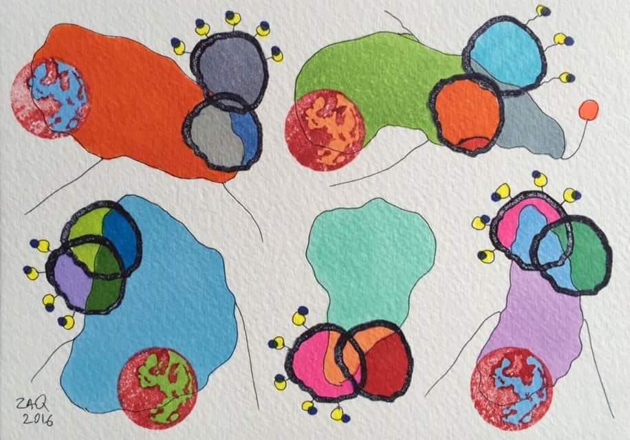
Like life, cells are subject to continuous change. Nothing in the vicinity of a cell remains still - unless death has interrupted its course. And the same goes for the inside of each cell. All sorts of molecules are being shuttled from one part to another, after having been created or on their way to being degraded. The cell membrane is also a very dynamic and supple structure, with molecules wandering through it constantly. One means of transport for shifting molecules around are known as endosomes. Endosomes are formed by the invagination of one part of the cell membrane - like a bubble budding towards the inside of the cell - which is then cleaved and able to float free in the cell's cytoplasm. Invagination then occurs on the surface of the endosomes themselves to form even smaller bubbles - or vesicles - that are in turn also set free. Like endosomes, these inner vesicles are a means of molecular transport too. It's a sort of Russian doll experience... The art of invagination per se may sound simple but it involves a lot of imagination on behalf of biology. Recently, scientists discovered a protein coined Snf7 that is providing hints as to how endosome invagination may occur, and the subsequent creation of vesicles.
In the same way we have buses, trains and planes to travel around the world, molecules have different ways of moving around a cell, or indeed from one cell to another. Endosomes are just one means of transport inside a cell and are made out of cell membrane, from where they bud. Imagine a cell, and that you're inside it. Now imagine a bubble budding from the membrane towards you, as though some invisible finger were pushing from the outside towards the inside. Now watch the bubble being severed from the membrane and left to float on its own. This is what an endosome is. And inside, it is carrying all the molecules - or cargo - that were part of the cell's membrane as it was being formed. Now, let's take it a little further: imagine sitting inside an endosome. Exactly the same chain of events occurs at the level of the endosome's membrane: a bubble buds, is severed, and floats off on its own. Bubbles that bud off the inside of endosomes are known as vesicles. And, like endosomes, they also transport molecules.
All kinds of proteins and protein complexes are involved in the making of endosomes and vesicles. Vesicle formation is driven by what are known as "endosomal sorting complexes required for transport", or ESCRTs. There are three ESCRTs (I, II and III) which, between them, relay cargo to where invagination will take place, promote invagination and then set the nascent vesicle free by severing it from the endosomal membrane. The ESCRT machinery and its role in budding were first observed in yeast for transporting molecules destined for degradation. However, it is now known that such budding events are similar to those used for HIV release and daughter cell abscission. Snf7 is the most abundant protein in the ESCRT-III complex and seems to be particularly involved in the budding process itself, i.e. membrane invagination.

Snf7 has a highly structured core domain of four alpha-helices, with a C-terminal alpha-helix that folds back onto the core domain. When the C-terminal end unfolds, Snf7 adopts an elongated "open" form that promotes protein-protein interactions; this is believed to be at the heart of membrane budding. What happens is Snf7 monomers can spontaneously fold into a single closed ring - or nucleation ring - which will, at one point, open. The "open form" induces Snf7b polymerisation; concomitantly, Snf7 is bent into a curve which is not "natural". When a second Snf7 monomer is added, it is also bent into an unnatural curve and so on. As the filament grows, the geometrical shape that eventually emerges is a two-dimensional spiral filament of Snf7 monomers on the cytoplasmic side of the endosome. Moreover, since the curvature of each open Snf7 monomer is unnatural, there is a mechanical stress which is heightened as the spiral filament elongates. And mechanical stress spells locked-up energy - in this case elastic energy.
There comes a point when the spiral stops growing - which seems to happen once the filament's length has reached 260nm. Why, no one really knows. Perhaps other proteins form a sort of wall in which it is confined, perhaps it reaches an energy threshold. Nevertheless, literally bursting with elastic energy, the spiral is thought to release its tension by lunging towards the inside of the endosome - in the manner of a jack in the box - thus forming a superhelix which drags the membrane along with it, forming the nascent vesicle. In this way, and to cut a long story short, Snf7 has the power to deform membranes by compressing elastic energy in a two-dimensional spring which ultimately expands.
It took molecular biology, mathematics, physics, chemistry and structural biology to imagine and test such a hypothesis, which seems to have convinced the scientific community. Snf7 filaments do indeed form spiral springs on endosome membranes and, upon some form of external cue, they are expected to release their stored elastic energy to form the budding vesicles. This demonstrates that a protein - depending on its conformation - has the ability to distort part of a membrane. Growing spirals no doubt speckle the endosome surface ultimately giving rise to vesicles, while similar invaginations occur at the level of the cell membrane. In this way, molecules are entering, travelling within and leaving the cell continuously. However, though Snf7 is proving to be essential for membrane invagination, it is also involved in membrane scission with the rest of the ESCRT complex - and membrane scission is an activity scientists are also very eager to understand at the molecular level.
